This is an acute inflammation of the lung parenchyma (lung tissue) caused by infectious agents or chemical irritations, leading to consolidation and exudation of the affected part.
Incidence
Pneumonia affects people world-wide. All age groups are affected, but is more common among the elderly and infants. Studies have shown that, 25% of boys are affected more than girls, also people in low social class, smokers and preterm babies have high incidence of pneumonia in early childhood. It is again common among the alcoholics and more common in winter and spring, but can occur in any season. The ratio of incidence among people living in Urban an Rural area is 2:1.
MODE OF TRANSMISSION
Pneumonia may be transmitted through droplet infection. It may also arise from aspiration of endogenous fluid by patients whose resistance is low or those on radiotherapy. It may again occur when the infections are carried to the lung by the blood stream.
PREDISPOSING FACTORS
The risk factors of pneumonia are as follows;
Lowered resistance of the host due to immunosuppressive drugs and diseases for example corticosteroid and AIDS respectively.
i. Cardiac Failure
ii. Chronic illness, for instance, diabetes mellitus
iii. Prolonged immobilization of patients
iv. Depressed cough reflex as seen in most unconscious patients
v. Exposure to extreme cold conditions
vi. Preterm babies
vii. Alcoholism and malnutrition
viii. Aspiration of infected mucus from the nose or sinuses, following simple infection of the upper respiratory tract.
ix. People who smoke.
AETIOLOGY
Many organisms can cause pneumonia, including bacteria, viruses and fungi, non-organic causes can also result in pneumonia. The bacteria causing pheumonia are;
Staphylococcus aureus
Streptococcus pyogens
Streptococcus pneumoia
Klebsiella pneumonia
Myocoplasma Pneumonia
Escherichia coli (E.coli)
Psudomonas aeruginosa
Proteus species
Bacteroid species
Haemophilus species
Pneumocysticcarini - Consistent with AIDS patients
Fungi and Virus
PATHOPHYSIOLOGY
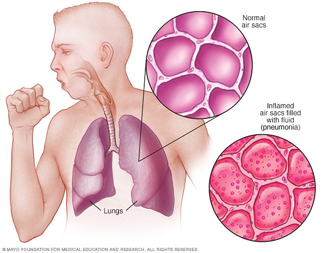
When the causative agent gets into the alveoli of the lungs, there is inflamatory process. This leads to exudates formation which cause oedema of the alveoli. The oedome create an optimum medium to bacteria growth and aid in spreading the infection to adjacent part of the lungs. The lung involved then undergoes consolidation. This consolidation is as a result of the lung with exudates. This leads to area of the lung to become inadequately ventilated resulting to inadequate oxygenation of the various blood coming into the lungs and eventually arterial hypoxia. The affected tissue becomes necrotic, the presence of the necrotic tissue then stimulates the individual to cough to bring foreign bodies out of the lungs.
CLASSIFICATION OF PNEUMONIA
A. Classification According to Morphology
Morphologically, pneumonias are recognized as;
Lobar Pneumonia; this is the type of pneumonia which large segments of one or more pulmonary lobes are involved. In this type, the entire lobe is affected, but lobular when part of the lobe is involved.
Bronchopneumonia; this is the type of pneumonia which begins at the distal or terminal bronchioles that become dogged with mucopurulent exudates.
Interstitials Pneumonia; in this, the inflammatory process is more or less confined to the alveoli walls (interstitium) and the peribronchial and interlobular tissues.
B. Classification According to Aetiology.
Bacterial/Pneumocystic Pneumonia
Viral Pneumonia
Aspiration Pneumonia
Fungal Pneumonia
Hypostatic Pneumonia (pneumonia caused by being bed ridden for a long time).
CLINICAL FEATURES
There is sudden onset of chills and fever.
i. Grunting respiration.
ii. Stabbing chest pains which is aggravated by coughing and respiration.
iii. Non-productive cough initially, but later productive cough with blood stains or yellowish sputum.
iv. Malaise and muscle pains.
v. Flashed cheeks, lip and nail beds cyanosed.
vi. Sore throat.
vii. There may be convulsions in infants.
viii. Abdominal distension in infants.
ix. There may be delirium in severe attacks.
x. There is high pulse rate.
xi. There is profuse sweating.
DIAGNOSIS
The following investigations are carried out to confirm the diagnosis;
i. Chest x-ray.
ii. Sputum for culture and sensitivity test.
iii. Physical examination.
iv. History taking, especially, of recent respiratory tract infection.
v. Blood examination, for example erythrocytes' sedimentation rate (ESR)
vi. Bronchoscopy
MANAGEMENT
Medical Management
Antibiotics such as ceftriaxone, ampicillin, crystalline penicillin.
Nursing Management
1. Nurse in upright position to assist pulmonary ventilation.
2. Give oxygen if there is cynosis.
3. Give drugs as ordered.
4. Educate client on predisposing factors.
5. Let patient lie on affected side of the lung (helps to splint the area and reduce pain when coughing).
COMPLICATIONS
1. Pleural effusion
2. Emphysema
3. Lung abscess
4. Cardiac failure
5. Meningitis, Pericarditis and Bronchiectasis
Others include; Myocarditis, Mental confusion and delirium, Otitis Media.
Read Also


0 Comments